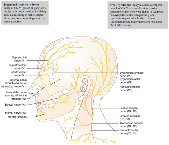
| Table 6-3 Sensory Innervation of Head and Neck |
| | Nerve Branch | | Sensory Innervation to: |
| V1: Ophthalmic branch |
| | Supratrochlear nerve | | Medial forehead, medial upper eyelid, frontal scalp |
| | Supraorbital nerve | | Most of forehead, portion of frontoparietal scalp, frontal sinus, upper eyelid |
| | Lacrimal nerve | | Lacrimal gland, conjunctivae, lateral eyelids | | | | | | | | | Reason why zoster lesions on tip of nose can be sign of ocular involvement (since both ciliary branch and external nasal branch come from nasociliary nerve) | | | | | | | |
|
| | External nasal branch of anterior ethmoidal (AE) nerve | | Nasal dorsum, tip, supratip, and columella | |
| | | | | | | | | | | | CN V → ophthalmic → nasociliary → AE nerve → external nasal branch | | | | | | | | |
|  |
| | Ciliary nerve | | Corneal surface |
| | | | | | | | | | | | CN V → ophthalmic → nasociliary → ciliary nerve | | | | | | | | | | | | | | |
|
| | Infratrochlear nerve | | Root of nose, upper lateral sidewalls , part of medial canthus, lacrimal sac |
| V2: Maxillary branch |
| | Infraorbital nerve | | Medial cheek, upper lip, lower nasal sidewall, nasal ala, lower eyelid |
| | Zygomaticofacial (ZMF) nerve | | Malar eminence of cheek |
| | Zygomaticotemporal (ZMT) nerve | | Temple and supratemporal scalp |
| | Superior alveolar and palatine nerve | | Upper teeth, palate, nasal mucosa, and gingiva |
| V3: Mandibular branch(both sensory and motor branches) |
| | Auriculotemporal nerve | | Anterior upper half of ear, tragus, preauricular cheek, anterior ½ of meatus, TMJ, external tympanic membrane, temple, temporoparietal scalp |
| | Buccal nerve | | Cheek, buccal mucosa, and gingiva |
| | Inferior alveolar | | Mandibular teeth |
| | Mental nerve | | Chin, lower lip |
| | Lingual nerve | | Anterior 2/3 of tongue (somatic sensation), floor of mouth, lower gingiva |
| Cervical plexus (C2–C4) |
| | Lesser occipital nerve C2 | | Neck and postauricular scalp, posterior upper half of ear |
| | Greater occipital nerve C2 | | Occipital scalp |
| | Transverse cervical nerve C2 and C3 | | Anterior neck |
| | Supraclavicular nerve C3 and C4 | | Anterior chest, clavicle, and shoulder |
| | Greater auricular nerve C2 and C3 | | Lateral neck, angle of jaw , inferior lateral cheek, anterior/posterior lower half of ear (include ear lobule), mastoid process, and postauricular skin |
| Other sensory nerves |
| | Facial nerve | | CN VII → chorda tympani
CN VII → small branches
(minor role in sensory) | | Taste sensation (anterior 2/3 tongue via chorda tympani), small portion of auditory meatus, concha bowl (variably innervated by branches of vagus and facial nerves), soft palate, pharynx |
| | Auricular branch of vagus nerve (CN X) | | CN X → auricular branch | | Posterior ½ of tympanic membrane and posterior wall of external auditory meatus |
| | Glossopharyngeal (CN IX) | | Taste and somatic sensation to posterior 1/3 of tongue |
| | | | |
