|
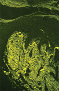
| Fig. 16.1 Lichen amyloidosis. Thioflavin-T demonstrates strong staining of fluorescent amyloid in the papillary dermis. (Courtesy of James E. Fitzpatrick, MD.) |
With light microscopy, amyloid appears as amorphous, hyaline-like, eosinophilic deposits. Amyloid demonstrates green birefringence with the alkaline Congo red stain, reddish metachromasia with crystal violet, and yellow-green fluorescence with thioflavin-T stain (Fig. 16-1). These stains are not absolutely specific for amyloid, as false-positive results may occur with the other hyaline-like deposition disorders.
